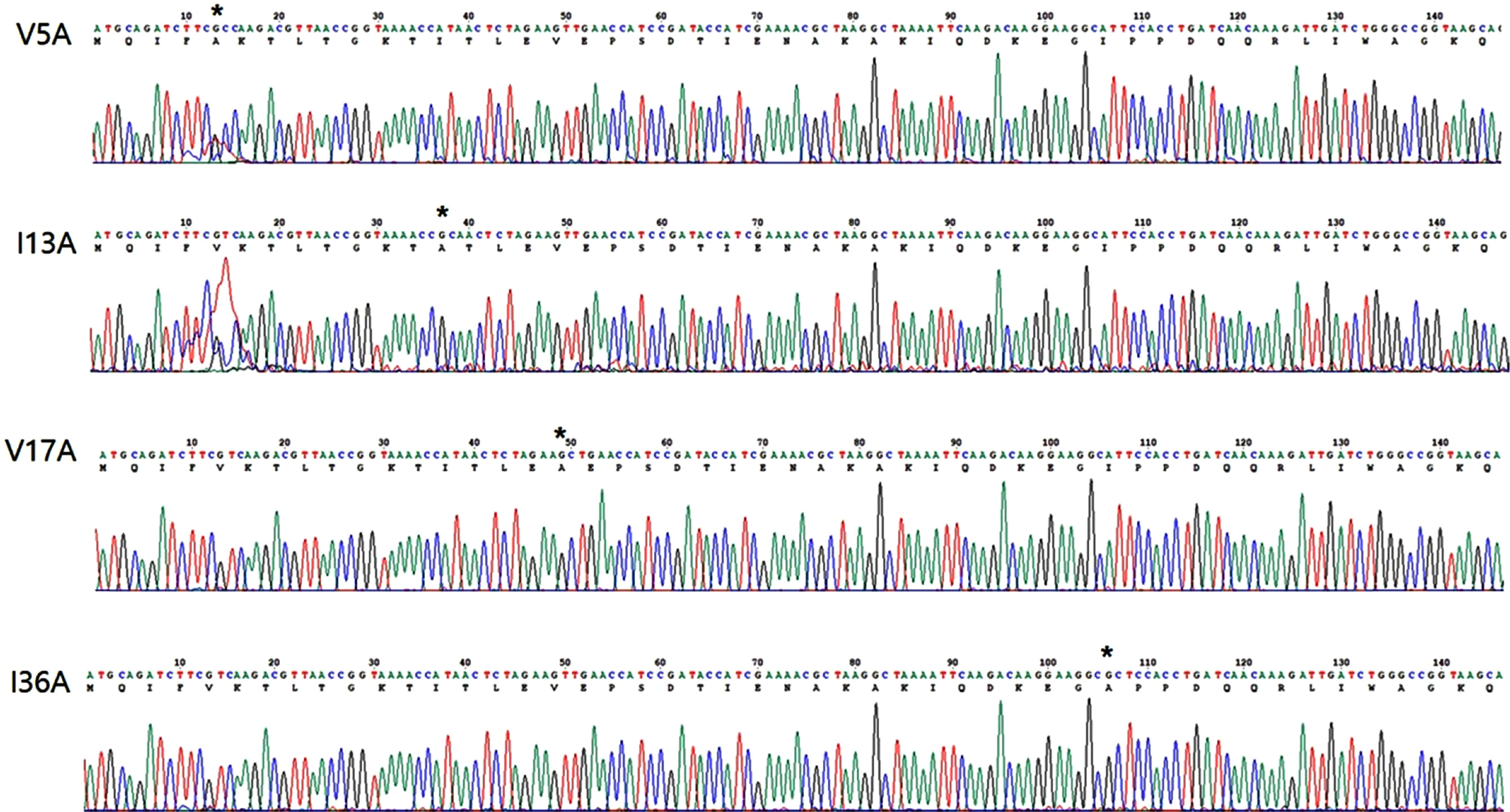
FigureS1.
DNA sequence of V5A, I13A, V17A and I36A. Mutated residue is designated as asterisk.
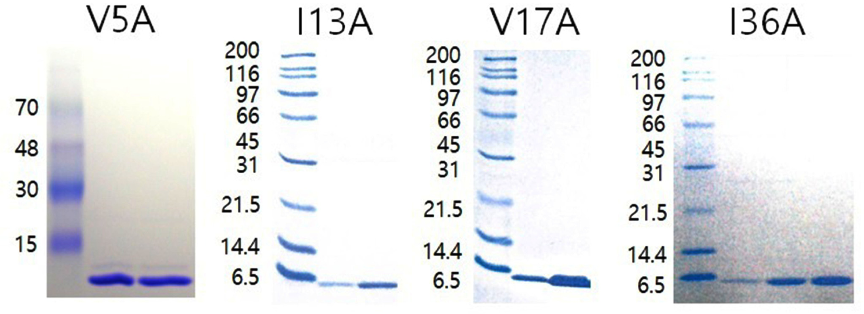
FigureS2.
SDS-PAGE of V5A, I13A, V17A and I36A. Gels were stained by coommassie brilliant blue. Purified mutant proteins were shown
as a single band.
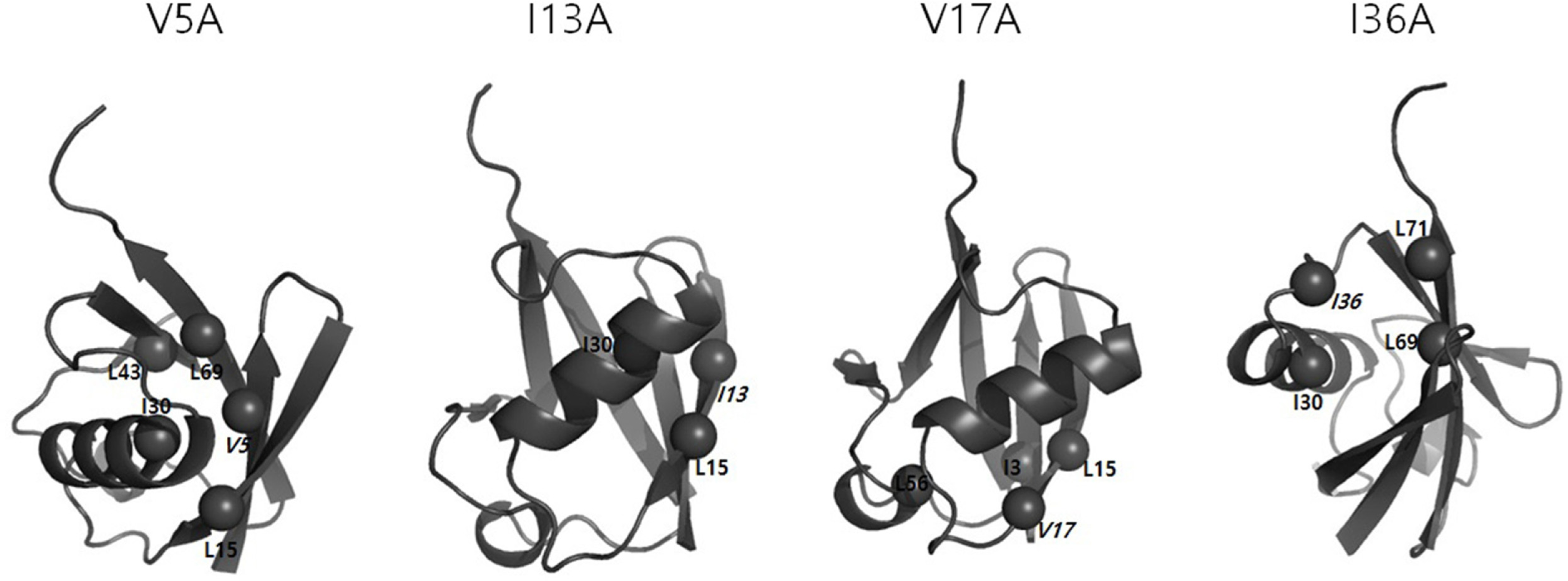
FigureS3.
Ribbon diagram of V5A, I13A, V17A and I36A. α-Carbon of mutated residue is shown as sphere and labeled in italic letter. α-Carbons
of residues that are located within 1.8 Å of mutated residue are shown as spheres and labeled in normal letter.